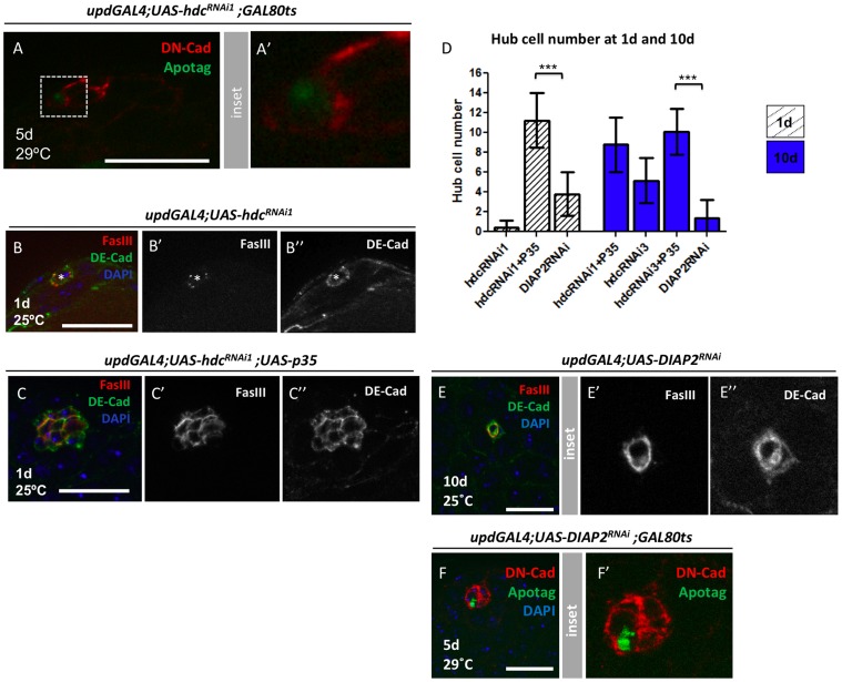
Figure 4. Loss of hub cells upon hdc reduction occurs via apoptosis.
(A) Example of an apoptotic hub cell found after RNAi-mediated reduction in hdc. DN-cadherin (red), Apotag (green), Genotype: updGal4;UAS-hdcRNAi1;Gal80ts. (B) Example of a 1d updGal4;UAS-hdcRNAi1 testis containing a residual hub composed of a single hub cell (*), Hub stained with both FasIII (red) and DE-cadherin (green), DAPI (DNA, blue). (C) Co-expression of p35 rescues the strong phenotype observed with hdcRNAi1. Compare to (B); (D) Hub cell number quantifications of different genotypes/time points; Mean and SD are shown; Welch’s t-test (***P<0.001). (E) Example of a compromised 10d updGal4;UAS-DIAP2RNAi testis; Note that DE-Cad shows two hub cells but one of them already lost FasIII staining); (F) Apotag positive hub cell in a updGal4;UAS-DIAP2RNAi testis; Scale bars 20 µm.

