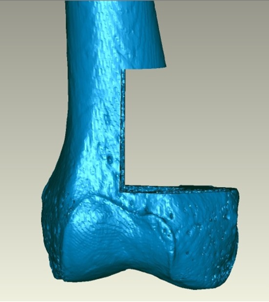
Fig. 1.

This is a three-dimensionally reconstructed CT image of a cadaveric femur with the distances from visible and/or palpable external landmarks depicted. The image was printed out for each manual resection specimen and used by the surgeon to perform the resection.
