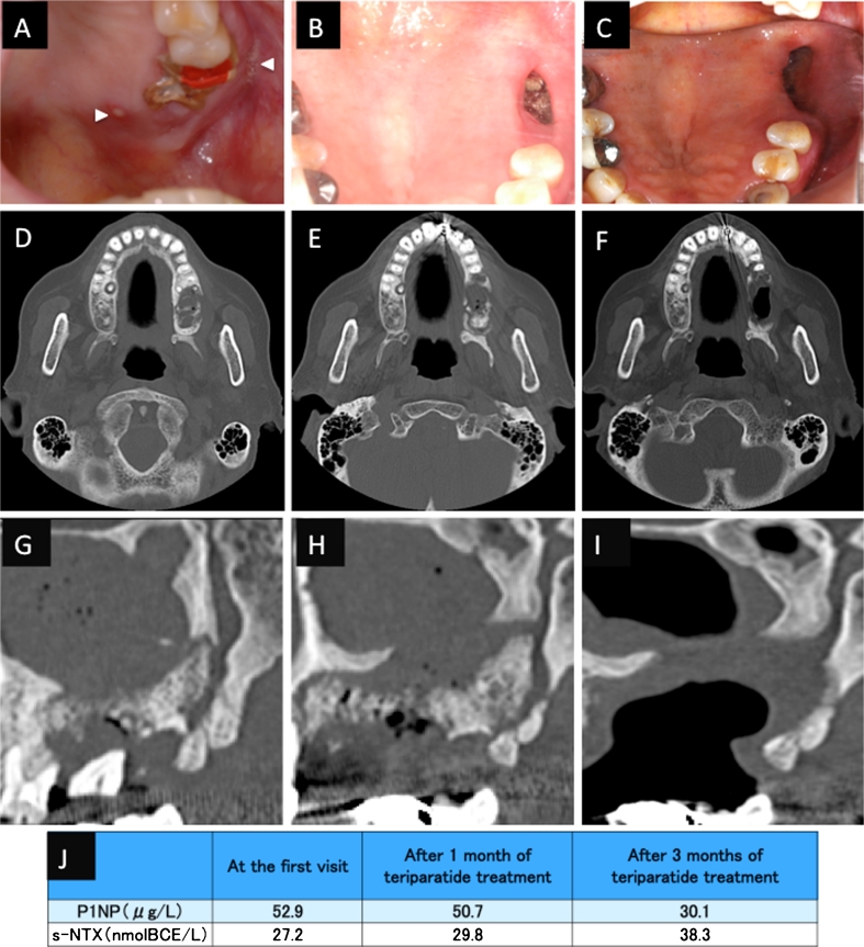
Fig. 1.
Case 1. An 87-year-old Japanese woman with a 4-year history of alendronate therapy. a At presentation, there were multiple fistulas with purulent discharge over the left maxillary ridge (arrowheads). After 3 months of conservative therapy, the unhealed wound was surgically debrided, and two teeth were extracted. b After 12 months of conservative treatment, there was still exposed bone in the upper jaw. c After 10 weeks of teriparatide treatment, the necrotic bone had healed, and there was complete soft tissue coverage of the intraoral wound. d, g Computed tomography (CT) images showing the maxilla before tooth extraction and debridement. e, h CT images after 1 year of conservative treatment, showing expansion of the BRONJ area. f, i CT images after 10 weeks teriparatide treatment, showing improvement of the maxillary sinusitis. j Levels of serum N-telopeptide of type I collagen (s-NTX) and serum N-terminal propeptide of type I collagen (P1NP)

