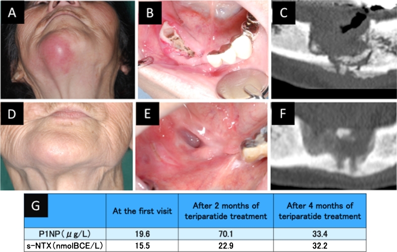
Fig. 2.
Case 2. An 87-year-old Japanese woman with a 4-year history of alendronate therapy. a External view showing submental redness. b Intraoral view showing exposed bone after the teeth were lost. c CT image at presentation. d External view after 3 months of teriparatide treatment. e Intraoral view after 2 months of teriparatide treatment, showing that the necrotic bone has healed and the defect is covered with normal mucosa. f CT image after 3 months of teriparatide treatment. g Levels of serum N-telopeptide of type I collagen (s-NTX) and serum N-terminal propeptide of type I collagen (P1NP)

