
Figure 8.
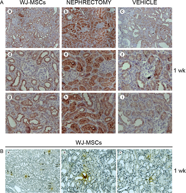
Hepatocyte growth factor (HGF) expression in injured kidney tissues at 1 week after intervention. (A) Representative micrographs illustrating HGF expression in injured kidney tissues. The overwhelming majority of positive staining resided in tubular cells. HGF staining in tubular cells was substantially intensified in cases of cell injection or nephrectomy. (a) through (c) Magnification ×200; (d) through (i) Magnification ×400. (B) Representative micrographs illustrating human HGF expression in injured kidney tissues. No positive-staining TECs were identifiable in kidney sections from sham-operated animals or IRI animals receiving vehicle injection (data not shown). By contrast, at 1 week after injection, positive staining (black arrows) was detected in kidney sections from cell-injected animals, mostly residing in cytoplasm of tubular cells. (a) Magnification ×100; (b) and (c) Magnification ×400.