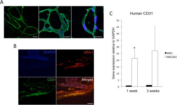
Figure 1.

Development of endothelial cell (EC) microvascular networks in a 3D copolymer scaffold. (A) Left: Light micrograph of GFP-expressing ECs organized in a microvascular network after 1 week of dynamic culture in vitro (20×). Scale bar = 50 μm. Middle: Confocal micrograph of ECs stained with human CD31 (green) organized in networks at 1 week in vivo (40×). Nuclei were stained with DAPI (blue). Scale bar = 20 μm. Right: Confocal micrograph of tubular CD31+ human EC at 1 week in vivo (60×). Scale bar = 10 μm. (B) Human CD31-positive cells were incorporated with surrounding UEA-1+ ECs from the mouse circulation after 1 week of implantation, as demonstrated by double staining (40×). Scale bar = 20 μm. (C) The relative expression of human CD31 was higher in MSC/EC constructs after 1 week of implantation, and the expression of human CD31 increased between 1 and 3 weeks. *P < 0.05; n = 6.
