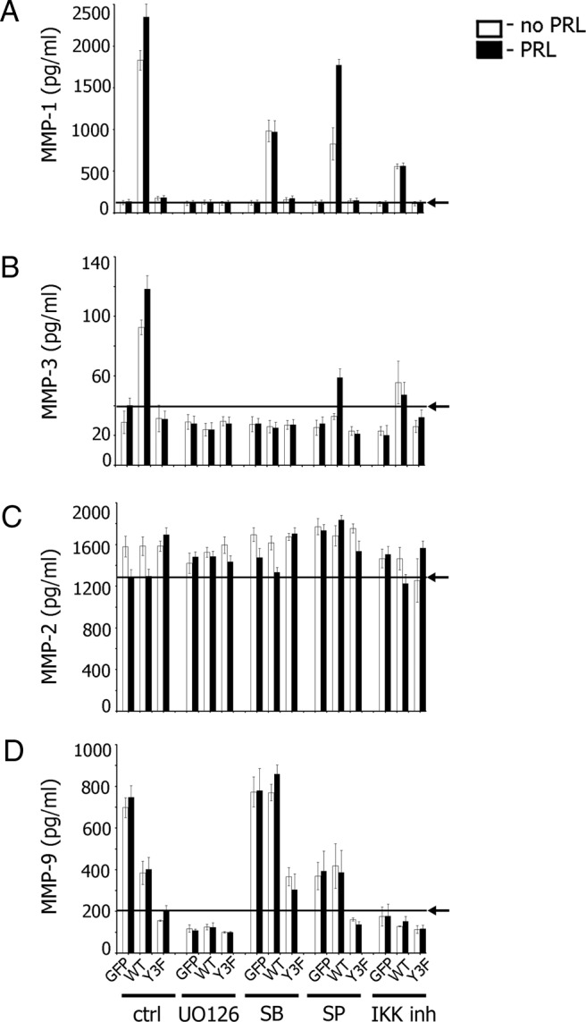
Figure 6.

Collagens and PRL-Dependent Tyrosyl Phosphorylation of PAK1 Regulate MMP Secretions via MAPKs and NFκB. TMX2–28 cells stably overexpressing GFP, PAK1 WT, or PAK1 Y3F were embedded in collagen IV (A–C) or in collagen I (D) and treated with inhibitors. For ERK1/2 inhibition, cells were deprived for 24 hours and treated with UO 126 (10 μM) for 1 hour prior to plating with or without PRL for 48 hours in the presence of UO 126 (final concentration, 10 μM). For p38 inhibition, cells were deprived for 24 hours, treated with 10 μM SB 203580 for 6 hours prior to plating the cells with or without PRL for 48 hours in the presence of SB 203580 (final concentration, 10 μM). For JNK1/2 inhibition, cells were deprived for 24 hours and treated with inhibitor (30 μM) for 1 hour prior to plating with or without PRL for 48 hours in the presence of SP 600125 (final concentration, 15 μM). For IKK inhibition, cells were deprived for 8 hours, followed by overnight incubation with IKK inhibitor VII (5 μM). Cells were plated the next day and incubated with or without PRL for 48 hours in the presence of IKK inhibitor VII (final concentration, 0.5 μM). The conditioned medium was assessed for secreted MMP-1 (A), MMP-3 (B), MMP-2 (C), and MMP-9 (D) by ELISA. Arrows indicate the average level of MMP secretion obtained in control GFP (A–C) or PAK1 Y3F (D) expressing cells treated with PRL. Bars represent mean ± SE. Each experiment was repeated 3 times.
