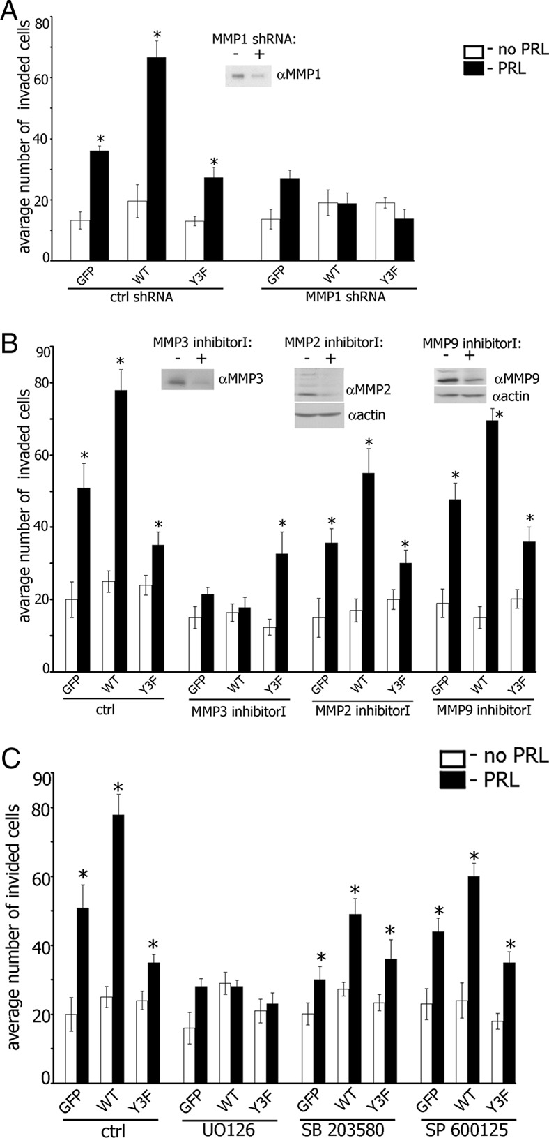
Figure 7.

Maximal Invasion of TMX2–28 Cells in Response to PRL Requires MMPs 1, 2, 3, and 9 and MAPK Activities. A, TMX2–28 cells stably overexpressing GFP, PAK1 WT, or PAK1 Y3F were transfected with control or MMP-1 shRNA and assessed for invasion as in Figure 2. Silencing efficiency was judged by immunoblotting of conditioned medium with anti-MMP1 Ab 48 hours after transfection. B, TMX2–28 cells stably overexpressing GFP, PAK1 WT, or PAK1 Y3F were treated with either MMP-3 inhibitor I (50 μM; overnight), MMP-2 inhibitor I (125 μM; overnight) or MMP-9 inhibitor I (50 μM; overnight) and assessed for invasion as in Figure 2. The conditioned medium (for MMP-3 inhibitor I) or cell lysates (for MMP-2 inhibitor I and MMP-9 inhibitor I) were analyzed by immunoblotting with the indicated antibodies. Latent (upper band) and active (lower band) forms of MMP-2 and MMP-9 were down-regulated by MMP-2 inhibitor I and MMP-9 inhibitor I, respectively. The expression levels of actin were used as an internal control. C, TMX2–28 clones were treated with MAPK inhibitors as in Figure 6 and assessed for the invasion as in Figure 2. Bars represent mean ±/- SE. *, P < .05 compared with untreated cells. Each experiment was repeated 3 times.
