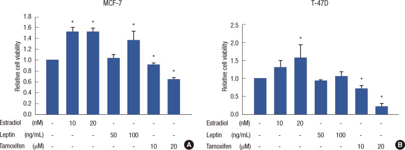
Figure 2.
Cell viability after single treatment of estrogen, leptin, and tamoxifen in MCF-7 and T-47D cells. MCF-7 cells at 70% confluence were synchronized in serum-free medium for 24 hours and treated. Experiments were performed at least 3 times. Cell viability assays are shown as histograms. The bars represent relative cell number (±standard deviation). Cell viability was assessed by CellTiter-Glo® Luminescent Cell Viability assay. (A) Estrogen and leptin had proliferative effects on MCF-7 cells compared to no treatment. Tamoxifen had a significant inhibitory effect on MCF-7 cell proliferation in a dose-dependent manner. (B) Estrogen had proliferative effects at 20 nM, and leptin had no significant effect on T-47D cell proliferation.
*Statistical significance compared to no treatment (p<0.05).

