Figure 10.
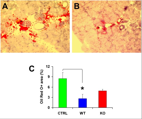
The Mst KO MDSCs are less effective than the WT MDSCs in reducing fat deposits in the injured mdx mouse gastrocnemius. (A) Representative picture of a positive field from frozen-tissue sections from the untreated mdx-injured gastrocnemius, adjacent to those shown in Figure 8D, fixed in formalin and stained with Oil Red O, showing mostly interstitial fat and occasional myofiber fat infiltration (200×). (B) Staining of a representative field from sections from the muscle implanted with WT MDSCs; the Mst KO pictures were similar, but the reduction in staining was less marked. (C) Quantitative image analysis of the tissue sections from the three rat groups, based on 12 fields per tissue section and the total positive area per section (percentage), calculated as a mean for three adjacent sections per rat, and five mdx mice/group. *P < 0.05. Mst KO, myostatin knockout; MDSC, muscle-derived stem cell; WT, wild type; mdx, X chromosome-linked muscular dystrophy.

