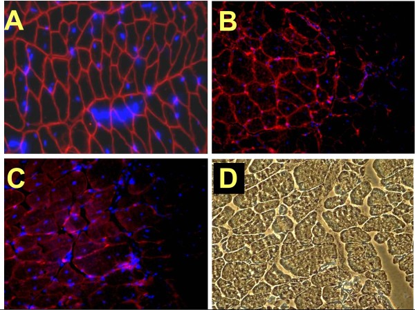
Figure 9.
The dystrophin+ MDSCs restore some dystrophin expression in the injured mdx gastrocnemius. (A) Myofibers from the intact gastrocnemius from the WT mouse, the source of WT MDSCs, show in a dual immunofluorescence merge all myofibers stained for dystrophin, and nuclei stained with DAPI (Vectashield mounting medium; Vector Laboratories, Burlingame, CA, USA) (200×). (B) In other tissue sections, DAPI-labeled implanted Mst KO MDSCs appear to have fused with the mdx myofibers, showing dystrophin+ staining in a small area. (C) A similar picture but with WT MDSCs. (D) The same field as in (C), examined under visible light, confirming the integrity of the myofibers, including the dystrophin- area. MDSC, muscle-derived stem cell; mdx, X chromosome-linked muscular dystrophy; WT, wild type; DAPI, 4', 6-diaminido-phenylindole; Mst KO, myostatin knockout.

