Figure 1.
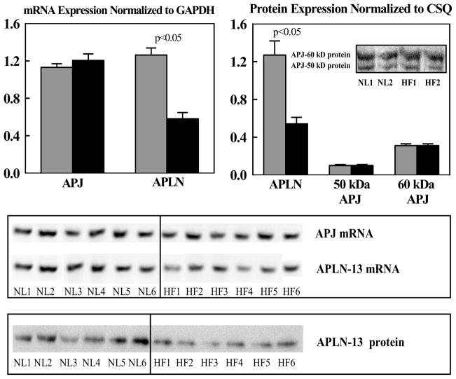
Top Left: Bar graph (mean ± SEM) depicting mRNA gene expression normalized to glyceraldehyde 3-phosphate dehydrogenase (GAPDH) for apelin-13 (APLN) and apelin receptor (APJ) in left ventricular myocardium of normal dogs (gray bars) and untreated dogs with coronary microembolization-induced heart failure (black bars). Top Right: Bar graph (mean ± SEM) depicting protein levels normalized to calsequestrin (CSQ) for apelin-13 (APLN) and apelin receptor (APJ) for bands at 50 kDa and 60 kDa in left ventricular myocardium of normal dogs (gray bars) and untreated dogs with coronary microembolization-induced heart failure. Insert shows sample of bands from Western blots in 2 normal (NL) dogs and 2 dogs with heart failure (HF). P-value denotes significant difference between normal dogs (gray bars) and heart failure dogs (black bars). Bottom: Immunoblots for mRNA and protein expression of apelin (APLN) and APJ receptor from all 6 normal (NL) and all 6 heart failure (HF) dogs reported in the study. P-value denotes significant difference between normal dogs and heart failure dogs (black bars).

