
Figure 2.
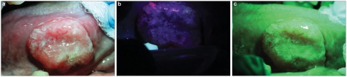
The use of Identafi optical system on a suspicious lesion showing loss of fluorescence and increase vascularity. (a) Picture of a non-healing ulcer on the right lateral border of the tongue of a 62-year-old male taken with the white reflectance (regular light) Identafi 3000 DentalEZ optical device. (b) Application of the Identafi DentalEZ device with violet fluorescent light shows dark area (loss of autoflurescence) in suspicious areas. (c) Application of the Identafi DentalEZ with the green amber reflectance light show increase vascularity in the suspicious areas. A biopsy taken from this area showed a moderately differentiated squamous cell carcinoma.