Figure 5.
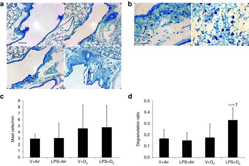
Effects of intra-amniotic LPS or vehicle plus postnatal air or hyperoxia exposure on mast cell accumulation and degranulation in hilar airways. (a) Toluidine blue–stained photomicrographs are shown. Top left: V+Air; top right: LPS+Air; bottom left: V+O2; bottom right: LPS+O2. Arrows indicate mast cells (not all the mast cells are indicated). Mast cells were located in the lamina propria, submucosa, and peribronchial adventitia and interstitium of hilar airways. (b) Degranulating mast cells in the hilar airways in the hyperoxia-exposed animals are indicated by arrows. Left: V+O2, right: LPS+O2. (c) Mast cell accumulation in the hilar airways was not significantly different among the treatment groups. (d) Mast cell degranulation ratio was significantly greater in the LPS+O2 group than in the other treatment groups. Original magnification: (a) ×100, (b) ×400. Bar: (a) 200 μm, (b) 50 μm. Data are the mean ± SD (eight per group). *P < 0.05 vs. V+Air group. **P < 0.05 vs. LPS+Air group. †P < 0.05 vs. V+O2 group. LPS, lipopolysaccharide; V, vehicle.

