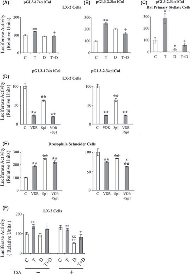
Fig. 2.
Effect of 1,25-(OH)2D3 (D) (10 nm) and TGFβS1 (T) (10 ng/ml) on the activity of (A) pGL-174 α1 and (B) pGL3-2.3k α1 in transfected LX-2 cells and (C) on the activity pGL3-2.3k α1 in transfected rat primary stellate cells. Effects of VDR (pcDNA-hVDR) and Sp1 (pPacSp1) expression vectors on the activities of (D) pGL-174 α1 and pGL3-2.3k α1 collagen promoters in transfected LX-2 cells and (E) in transfected Drosophila Schneider cells. (F) Effect of trichostatin on the activity of the pGL3-2.3k α1 collagen promoter in the presence of 1,25-(OH)2D3 and or TGFβ1. The cells were cultured for 24 h with or without trichostatin A (TSA) (100 nm), 1,25-(OH)2D3 (D) (10 nm) and TGFβ1 (T) (10 ng/ml). Luciferase activities are shown as percentages of the control value. The data are expressed as means ± of 6−8 determinations. *P < 0.05 vs. respective control. **P < 0.01 vs. respective control. +P < 0.05 vs. TGFβ1. ++P < 0.01 vs. TGFβ1. XP< 0.01 vs. VDR. XXP< 0.01 vs. TSA alone.

