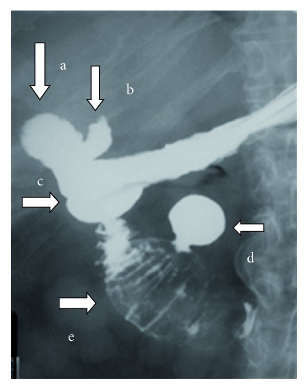
Figure 6.

Five minutes after intake of the oral contrast all the anatomic elements are well visualised: (a) the gallbladder filled with contrast, (b) the cystic duct filled with contrast, (c) the cholecystoduodenal fistula, (d) a duodenal diverticulum of the 3rd part filled with contrast, and (e) a large gallstone impacted on the 2nd to 3rd part of the duodenum.
