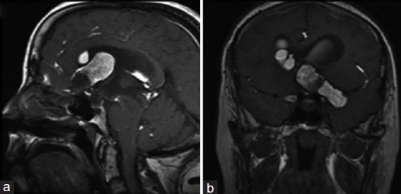
Figure 2.

Preoperative sagittal and coronal T1 MRI of the brain (a and b) shows a large T1 hyperintense multilobulated lesion extending along the floor of the left anterior and middle cranial fossa, with areas of T1 hypointense signal around the component in the left anterior cranial fossa. Locules of T1 hyperintense material are present throughout the subarachnoid spaces along the sylvian fissures bilaterally, extending into the frontal sulci, parasagittal frontal lobes, tectum, and into the posterior fossa. Fat-fluid levels are present within the ventricular system, with evidence of obstructive hydrocephalus
