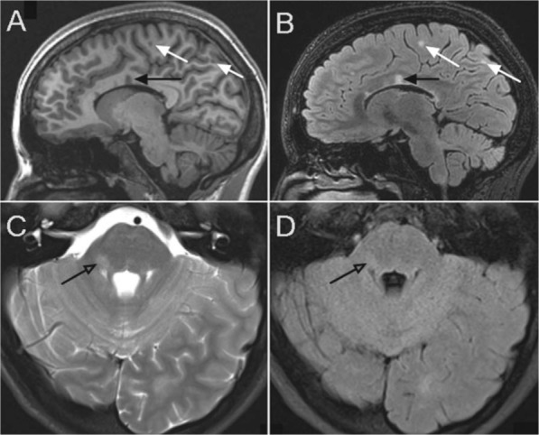
Figure 1.

Lesions in subject with relapsing remitting MS. Top images are sagittal images with corpus callosum lesions (black arrows) and juxtacortical lesions on the T1 weighted image (A) and T2 FLAIR weighted image (B). Bottom images clearly demonstrate the improved sensitivity of posterior fossa lesion detection (open arrow) on standard T2 weighted images (C) over T2 FLAIR (D).
