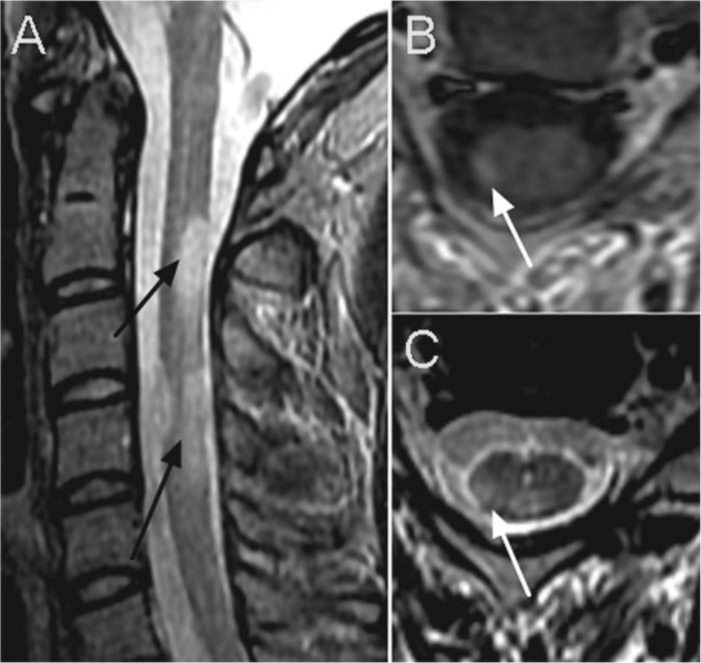
Figure 3.

Typical spinal cord lesions in a patient with RRMS. On the sagittal STIR sequence (A) multiple ovoid lesions (black arrows) are seen extending across one vertebral segment. The images on the right show axial cuts of the same individual, showing the typical pattern of ovoid lesion in the right dorsolateral aspect of the cervical spinal cord (white arrow). (B) post gadolinium contrast T1 weighted image, (C) T2 weighted image.
