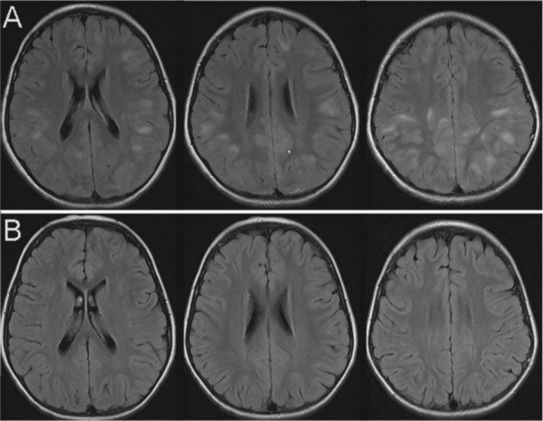
Figure 6.

Images of acute disseminated encephalomyelitis (ADEM) in a pediatric patient. The T2 FLAIR axial images were acquired during the symptomatic phase of the disease (A) with typical poorly demarcated lesions with white matter and grey matter involvement. There was no contrast enhancement of any of these lesions (not shown). On follow up imaging three months later (B) no residual signal change is noted.
