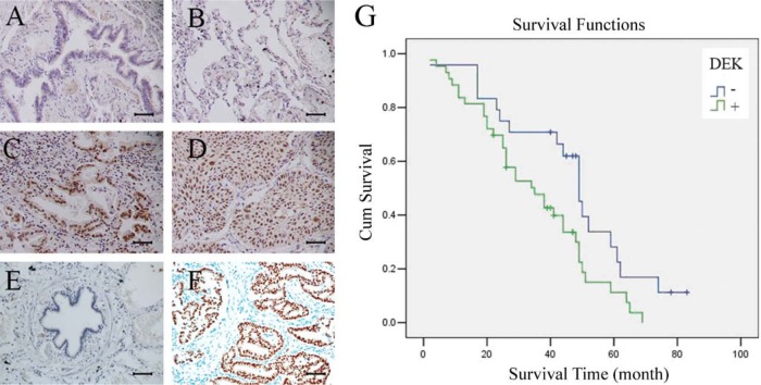
Figure 1.
DEK expression in non–small cell lung cancer (NSCLC) specimens. (A) Negative staining in normal bronchial epithelium in non-cancerous lung tissue. (B) Negative staining in normal pneumocytes in the alveoli of non-cancerous lung tissue. (C) Positive DEK staining in a case of adenocarcinoma. (D) Positive DEK staining in a case of squamous cell carcinoma. (E) Negative control. (F) Positive control (DEK is strongly positive in invasive ductal carcinoma of breast). Scale bars: 50 um. (G) Survival curves of patients with positive and negative DEK expression.

