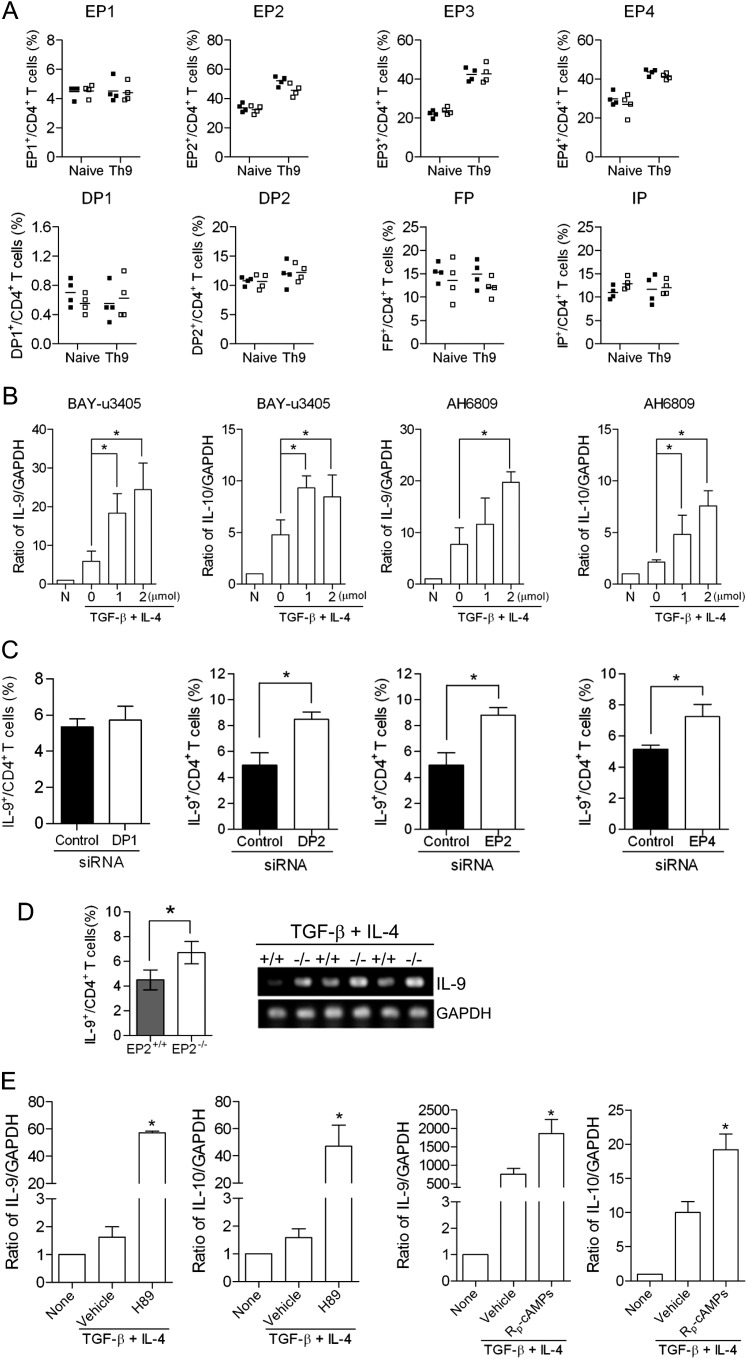
Figure 5.
Regulation of T helper cell type 9 (Th9) cell differentiation is mediated through prostanoid receptors. (A) The percentage of prostanoid receptor (EP1, EP2, EP3, EP4, DP1, DP2, FP, and IP)–positive naive CD4+ T cells and in vitro differentiated Th9 cells from cyclooxygenase (COX)-2+/+ and COX-2−/− mice was determined by flow cytometry. Lines indicate the mean, and each symbol (COX-2+/+, filled squares; COX-2−/−, open squares) represents an individual mouse. (B) The effects of the TP/DP2 receptor antagonist, BAY-u3405, or the EP1–3/DP1 receptor antagonist, AH6809, on Th9 differentiation of naive CD4+ T cells were investigated by real-time RT-PCR (n = 3; *P < 0.05 versus vehicle). (C) Naive CD4+ T cells from wild-type (WT) mice were transfected with DP1, DP2, EP2, or EP4 receptor siRNAs, or control siRNA. Transfected cells were then differentiated in the presence of anti-CD3, anti-CD28, anti–INF-γ, transforming growth factor (TGF)-β, and IL-4 for 5 days and the percentage of Th9 cells was analyzed by flow cytometry (n = 3; *P < 0.05 versus control siRNA). (D) Th9 cell differentiation of naive CD4+ T cells isolated from EP2 receptor knockout mice and WT controls were investigated by flow cytometry and RT-PCR (n = 5; *P < 0.05 versus WT). (E) Two different protein kinase A (PKA) inhibitors (H89 and Rp-cAMPs) significantly increase Th9 cell differentiation, as measured by IL-9 and IL-10 expression in vitro (n = 3; *P < 0.05 versus vehicle).

