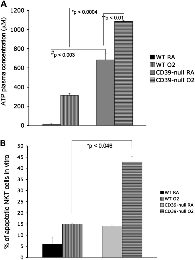
Figure 7.
Increased plasma ATP concentrations and iNKT apoptosis in CD39-null mice. (A) High-performance liquid chromatography analysis of plasma shows that ATP plasma concentrations were significantly increased in the CD39-null animals after oxygen exposure, compared with wild-type animals (P < 0.0004). All error bars represent the SEM (n = 3 per group). (B) In vitro apoptosis assay. iNKT cells from wild-type and CD39-null animals were cultured (104 cells per well) in room air (RA) or 95% O2/5% CO2 for 72 hours. Cells were then stained with PECy7-conjugated anti-NK1.1, FITC-conjugated anti-CD3, and APC-conjugated anti–annexin V and propidium iodide (PI). Apoptotic cells were defined as annexin V+/PI− and annexin V+/PI+. CD39-null iNKT cells showed significantly higher levels of iNKT cell apoptosis under oxygen than did the wild-type iNKT cells (P < 0.046). All error bars represent the SEM (n = 2 per group).

