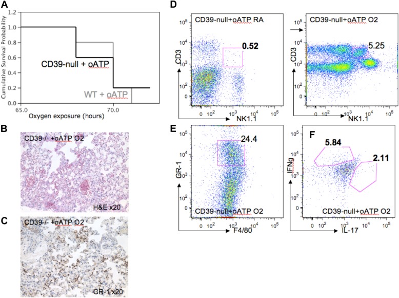
Figure 8.
P2X7 receptor antagonism induces hyperoxic lung injury in CD39-null mice. The P2X7 antagonist oxidized ATP (oATP) was administered to wild-type and CD39-null animals. Mice were then exposed to either room air or 100% oxygen for 72 hours (n = 3 per group). (A) Kaplan-Meier survival curves show that survival in CD39-null animals treated with oATP decreases dramatically, and approaches survival rates of the wild-type mice after oATP treatment. (B) Hematoxylin-and-eosin staining of oxygen-exposed lungs reveals severe lung injury in the oATP-treated CD39-null mice, with prominent (C) GR-1+ PMN lung infiltrates. Pulmonary mononuclear cells were extracted and stained with PECy7-conjugated anti-NK1.1, FITC-conjugated anti-CD3, PE-conjugated anti–GR-1, and FITC-conjugated anti-F4/80. (D) Flow cytometry analysis (FACS) confirmed significant increases in the pulmonary iNKT cell population in oATP-treated animals (5.2% of total pulmonary mononuclear cells), compared with the untreated CD39-null mice (Figure 3C) (1.75% of total mononuclear cells). All error bars represent the SEM (n = 3 per group). (E) Pulmonary polymorphonuclear cell (PMN) infiltrates were identified in the oATP-treated CD39-null animals. (F) Intracellular staining using flow cytometry (FACS) from pulmonary-derived iNKT cells. Mononuclear cells were extracted from oxygen-exposed lungs and stained with PECy7-conjugated anti-NK1.1, FITC-conjugated anti-CD3, PE-conjugated anti–IL-17, and Pacific blue–conjugated anti–IFN-γ. NK1.1+/CD3+ cells were assessed for IL-17 and IFN-γ expression. (F) oATP-treated CD39-null mice produced higher concentrations of IL-17 and IFN-γ, compared with (Figure 4D) untreated CD39-null cells after 100% oxygen exposure.

