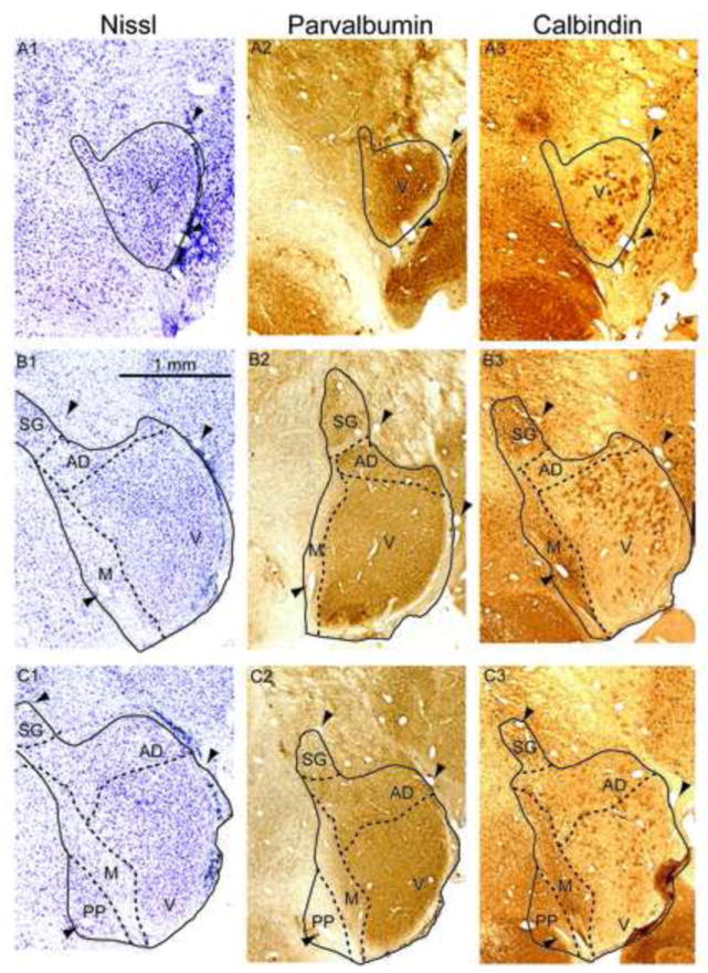
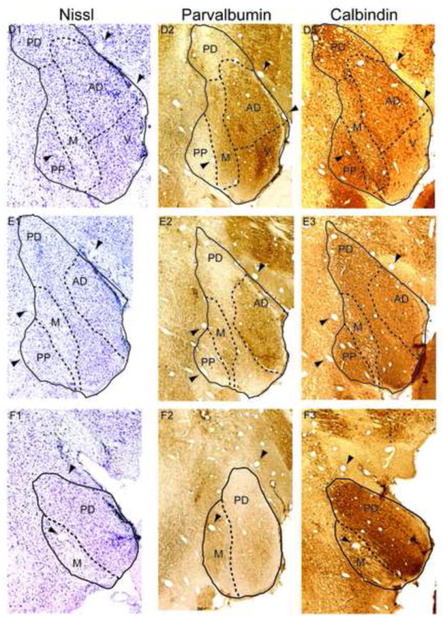
Figure 1. Marmoset MGB and calcium binding proteins.


Left column: Nissl stain, with MGB subdivisions indicated by dashed lines. Arrowheads indicate blood vessels that are found in all three adjacent sections in a row. Middle column: Parvalbumin immunostaining. Right column: Calbindin immunostaining. The topmost row (A1-A3) is the most rostral portion of MGB. Subsequent rows progress caudally by approximately 240 m per row. Abbreviations: V, ventral division, PD, posterodorsal division, AD, anterodorsal division, M, medial division, SG, suprageniculate nucleus, PP, peripeduncular region.
