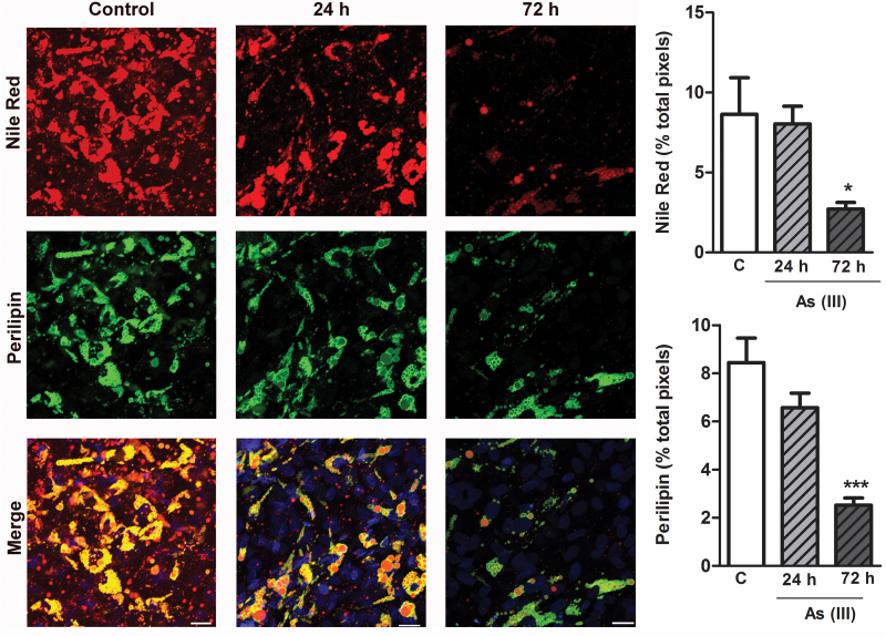
Fig. 4.
As(III) causes progressive loss of adipocyte PLIN1-coated lipid droplets. Adipocytes were differentiated and grown on glass coverslips and then treated with 1μM As(III) for the indicated times. The cells were fixed and stained for neutral lipid droplets (Nile Red), PLIN1 (green), and nuclei (DAPI blue) content. Images of four fields per cover slip were captured at ×40 (scale bars = 50 μm) and the thresholded fluorescence quantified and averaged. The data in the graphs present mean ± SEM percentage of positive pixels normalized to DAPI staining in four separate cultures representative of two separate experiments. Data were analyzed by ANOVA and Newman-Keuls post hoc test for differences between groups (*p < 0.05 and ***p < 0.001 from control).

