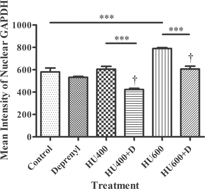
Fig. 1.
Analysis of the confocal images of GAPDH immunofluorescence in the lumbosacral regions of GD 9 embryos using IMARIS. The immunofluorescence intensity of isolated nuclear GAPDH is represented here. HU 400, 400mg/kg hydroxyurea; HU600, 600mg/kg hydroxyurea; D, deprenyl. Two-way ANOVA and a post hoc Bonferroni correction were done. Asterisks (***) denote a statistically significant difference (p < 0.001). † denotes a significant difference between the hydroxyurea-treated groups in the absence and presence of deprenyl (p < 0.05).

