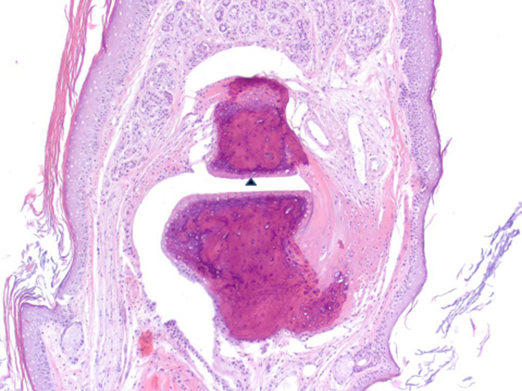
Figure 10.
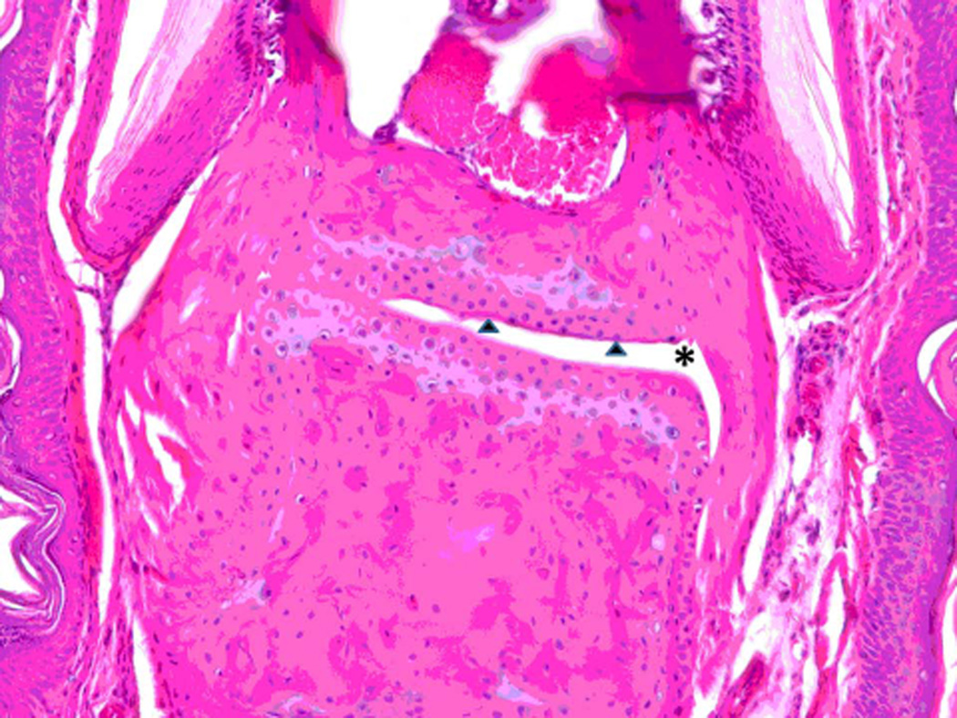
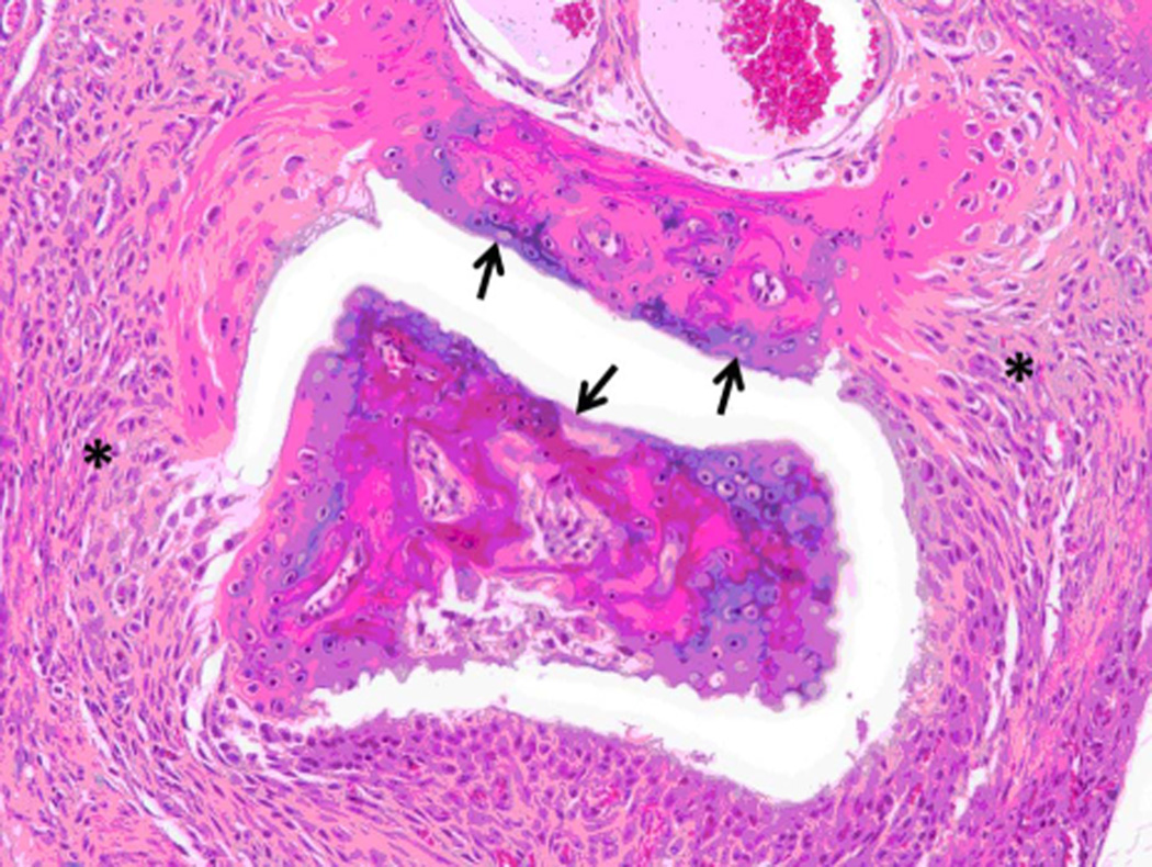
Histopathology analysis of sample from paws from normal, arthritic, and treated DBA/1 mice. Sections of paws from 6 mice were chosen, and representative sections were chosen for final analysis. A) Normal phalangeal joints of hind foot. Cartilaginous surfaces are intact and smooth (arrow heads) and joint space is clear (*). H & E staining. Original magnification 200X. B) Arthritis hind paw with severe (grade 4) arthritis of phalangeal joint. Joint is swollen. Marked cartilaginous erosion of joint surface and pannus circumscribing the phalanges. H & E staining. Original magnification 200X.
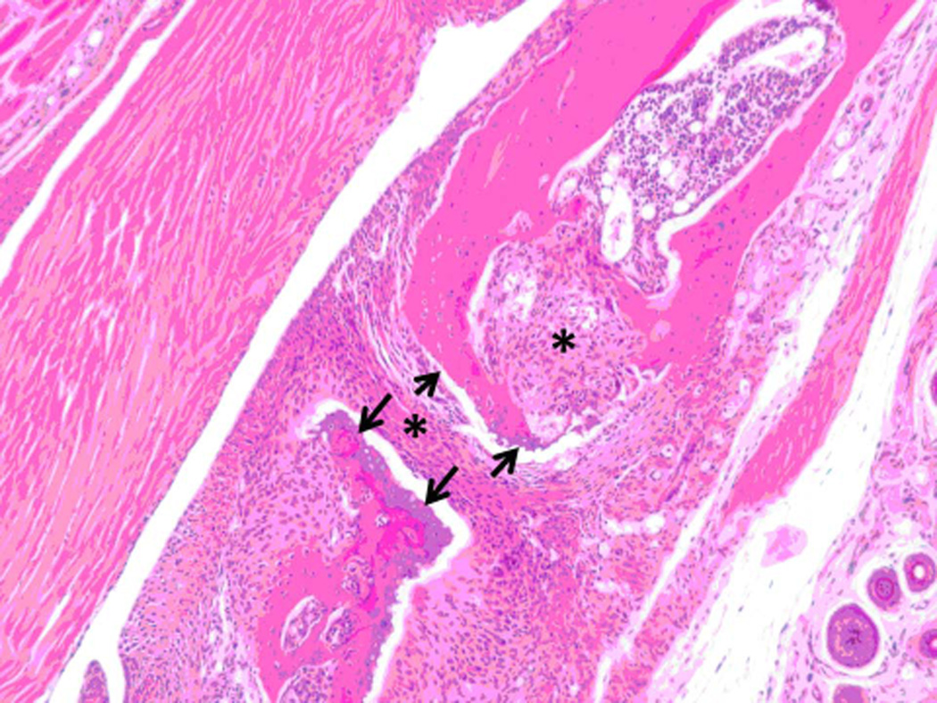
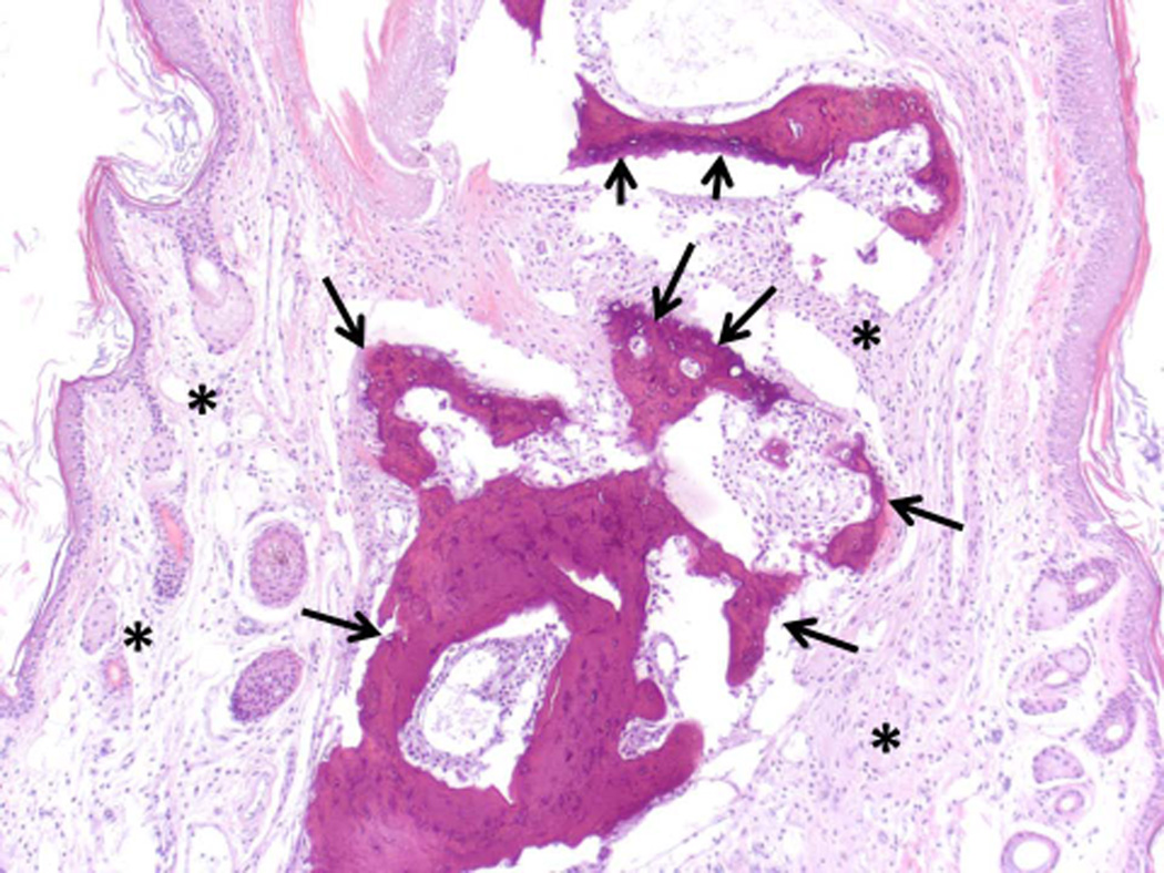
C) and D) Arthritis hind paw with severe (grade 4) erosion of cartilage and bone resorption (arrows) of phalanges. The connective tissue (*) of the dermis, hypodermis, and peristeum is mildly edematous and infiltrated with neutrophils, mixed mononuclear inflammatory cells, and fibroblasts. H & E staining. Original magnification 100X. E) Hind paw of mouse treated with peptide 6 (0.5 mg/kg). Joint space is clear (*) with normal cartilage interface. Connective tissue surrounding joint is only very minimally infiltrated with mixed inflammatory cells. H & E staining. Original magnification 100X.

