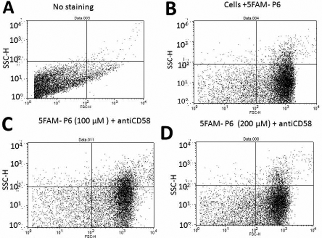
Figure 3.
Competitive binding of fluorescently labeled peptide 6 (5FAM-peptide 6) with anti-CD58 to Caco-2 cells expressing CD58 monitored by flow-cytometry. 10,000 cells were counted using green fluorescence. The figure represents forward and side scatter representation of Caco-2 cells. A) Caco-2 cells without peptide or antibody. B) Cells with 5FAM peptide 6, 100 µM. C) Cells with 5FAM-peptide 6, 100 µM, and anti-CD58. More than 75% of the cells were stained with peptide 6. D) Cells with 5FAM peptide 6, 200 µM, and anti-CD58. More than 80% of the cells were stained with peptide 6.

