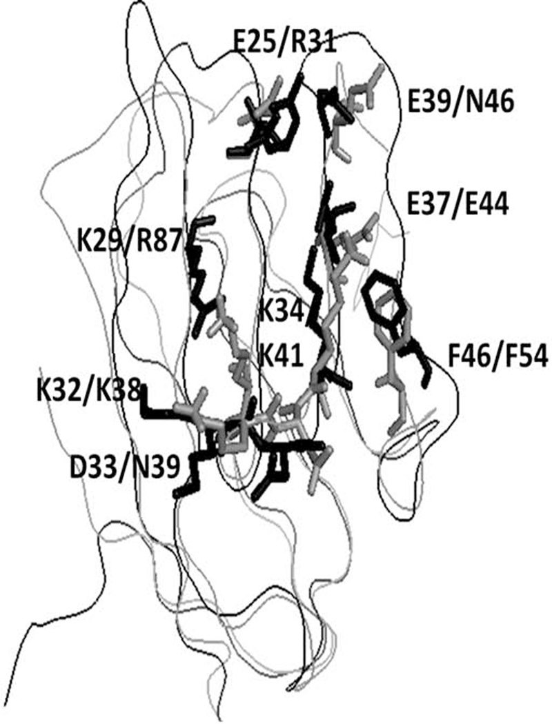
Figure 4.
Comparison of crystal structures of CD58 and CD48. CD58 is shown as gray and CD48 shown as black. Amino acids from CD58 that are shown to be important in binding to CD2 using mutagenesis are shown as sticks with labels. For comparison, amino acids of CD48 are also shown. Single letter codes for amino acids; first label refers to CD58 and second label refers to CD48.

