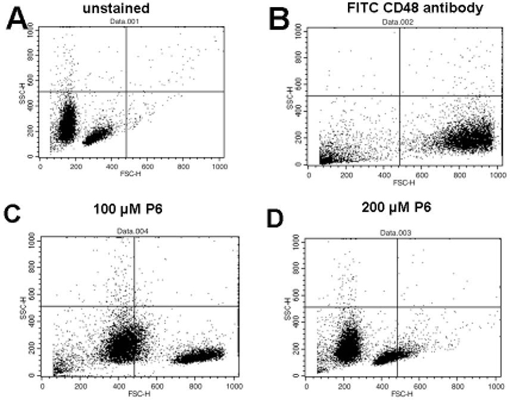
Figure 5.
T cells were collected from mice spleen and treated with FITC-labelled anti- CD48 along with peptide 6 at various concentrations to evaluate the competitive binding. A) unstained T-cells were observed in the lower left quadrant. B) Cells+ FITC-labelled anti-CD48; nearly 80% cells shifted into the lower right quadrant. C) Cells+ FITC-labelled anti-CD48+ peptide 6 100 µM. More than 50% cells were found to be unstained in the lower left quadrant. D) Cells+ FITC-labelled anti-CD48+ peptide 6 200 µM; more than 90% of cells in the lower left quadrant were found to be unstained.

