Figure 6.
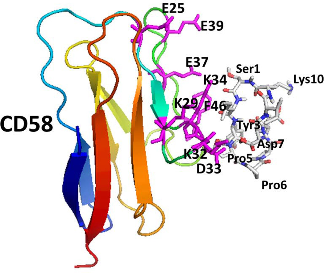
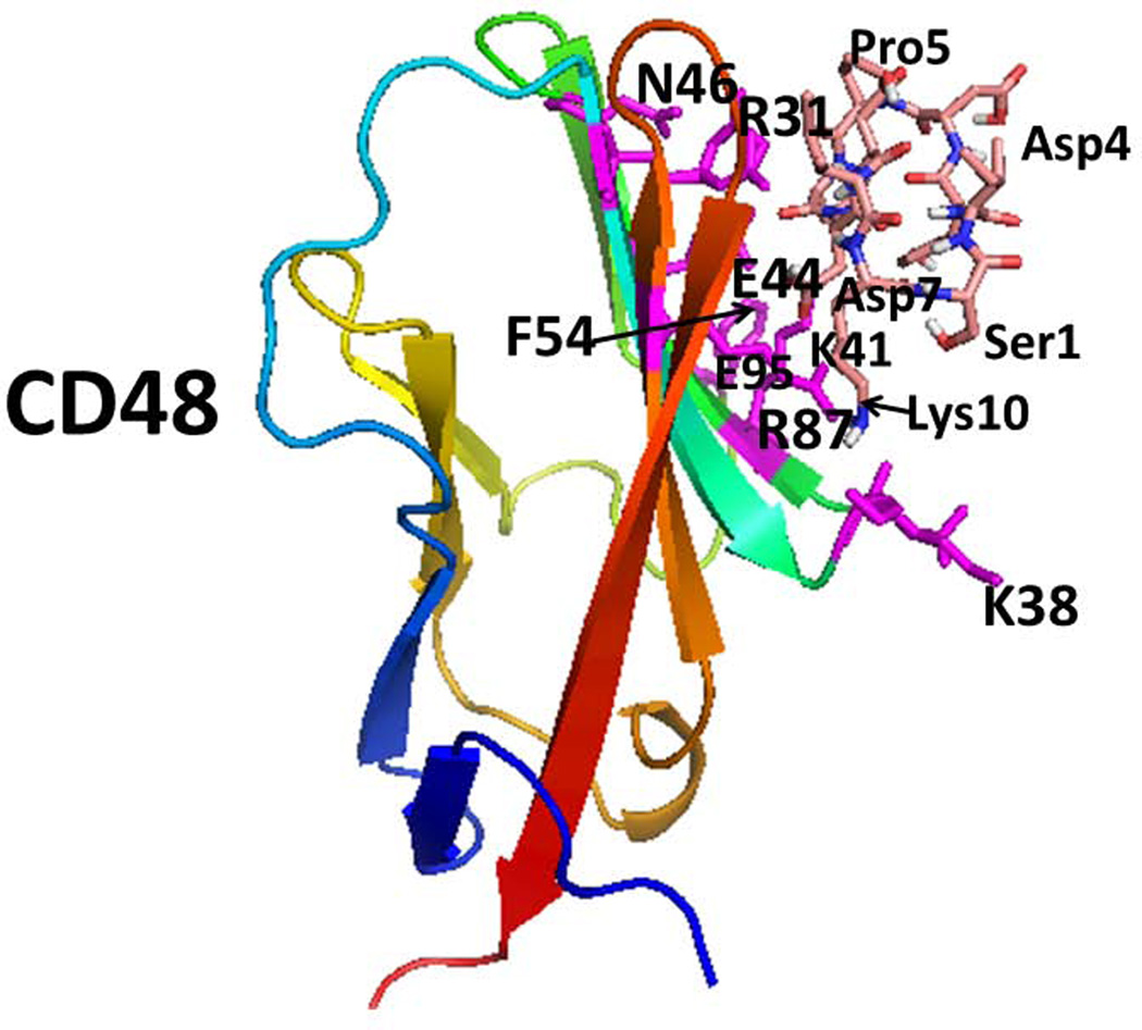
Proposed model for binding of peptide 6 on CD58 and CD48 using docking studies. A) Low energy docked structure of peptide 6 binding to adhesion domain of CD58. Amino acid residues that are shown to be important in binding to CD2 on CD58 are shown as sticks (magenta). Peptide 6 peptide is shown as sticks. B) Low energy docked structure of Peptide 6 binding to adhesion domain of CD48. Residues that are important in binding to CD2 were compared with CD2-CD58 structure. Similar residues in CD48 are represented as sticks (magenta). Amino acids from the protein are labeled with single letter codes and those from the peptide are labeled with three letter codes for clarity.


