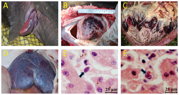
Figure 1.

Pathologic changes of elephant endotheliotropic herpesvirus (EEHV) –associated disease found during field necropsy. (A) Asian elephant (Elephas maximus) with cyanosis of the tongue attributed to EEHV disease; (B) epicardial surface of the heart (apex view) showing severe extensive hemorrhage; (C) ventricular endocardial surface of the heart showing multifocal areas of ecchymotic hemorrhage; (D) serosal membrane surface of the liver showing diffusely scattered petechial hemorrhage; (E,F) photomicrograph of two capillary endothelial cells containing typical basophilic intranuclear viral inclusion bodies from necropsy liver tissue. H&E stain, bar=25 μm.
