
Fig 2.
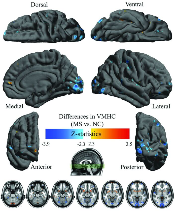
Comparison results of whole-brain voxelwise VMHC maps between patients with MS and healthy control participants (NC) showed regions with significantly decreased (blue) and increased VMHC (red and yellow) in patients compared with control participants corrected with Gaussian random field theory (minimum Z > 2.3; cluster level; P < .05, corrected).