
Figure 1.
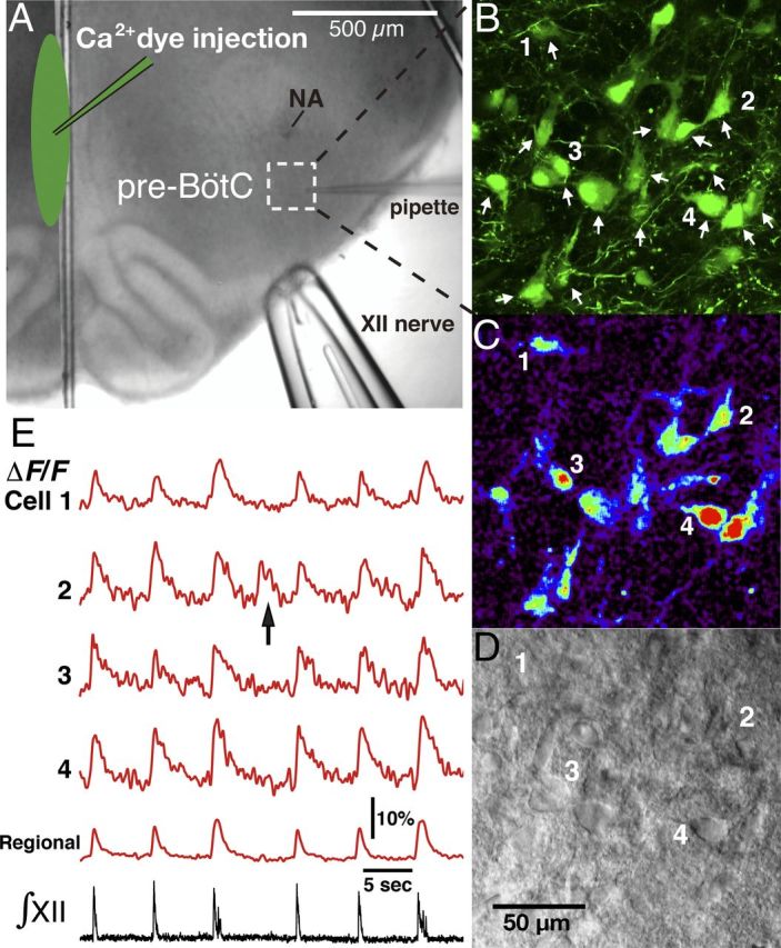
Overview of experimental in vitro neonatal rat rhythmic slice preparation with inspiratory neuronal activity imaging via Ca2+-sensitive fluorescent dye. A, Ca2+-sensitive fluorescent dyes (Ca2+ dyes) were injected along the midline of the slice (shown by infrared Dodt contrast structural image) to label pre-BötC neurons with contralaterally projecting axons. B, Illustrative example of OGB labeled neurons in the pre-BötC area marked by dashed square in A. Two-photon fluorescence, single optical section. Arrows indicate inspiratory neurons identified by analyzing their Ca2+ fluorescence signals in real time simultaneously with inspiratory hypoglossal nerve activity, which is used to monitor inspiratory network activity. Neurons labeled (1–4) are indicated in images and their fluorescence activity is shown in C and E. C, Background-subtracted image from B showing inspiratory activity-related “flash” of increased fluorescence intensity during inspiration. D, Dodt contrast image simultaneously captured with OGB fluorescent image in B and C, providing structural view of individual neurons 1–4 and other neurons that exhibited inspiratory fluorescence transients. E, Fluorescence intensity signals from cells 1–4 and from the captured pre-BötC region (spatially averaged fluorescence signal) showing Ca2+ transients synchronized with inspiratory hypoglossal nerve activities (∫XII). The arrow in cell 2 trace indicates an example of Ca2+ fluorescence transient associated with ectopic neuronal bursting independent of synchronized network activity as indicated by ∫XII activity.