
Figure 5.
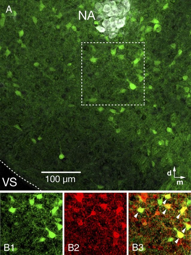
Distribution of GAD67-GFP positive neurons in the pre-BötC region and coexpression of glycine in GAD67-GFP transgenic mouse. A, GAD67-GFP positive neurons distributed within the pre-BötC (10× objective). Motoneurons in the NA were stained with ChAT antibody (white). B, Single optical plane images of GAD67-GFP positive (B1) and glycine (B2) immunolabeled neurons in the pre-BötC area marked by dashed box in A. Merged image (B3) shows a mixed population of pre-BötC inhibitory neurons, including GAD67-GFP positive neurons with glycine immunolabeling (yellow, arrowheads) or without glycine immunolabeling (green), and GAD67-GFP negative neurons with glycine immunolabeling (red). VS, Ventral surface.