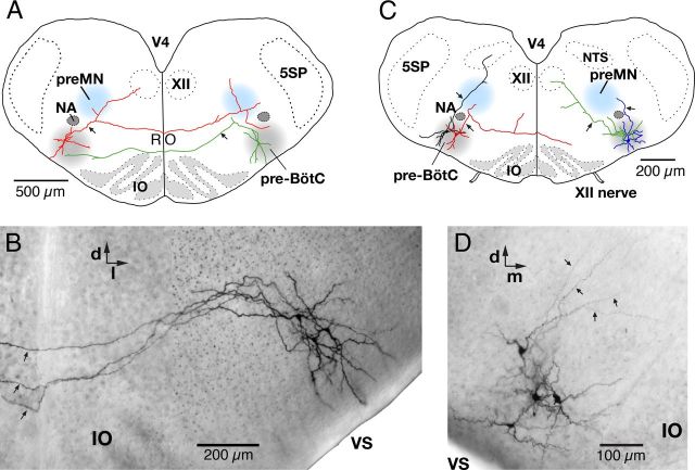
Figure 8.
Axonal projection patterns of pre-BötC inspiratory neurons. A, Representative examples of axonal projection patterns of pre-BötC inspiratory neurons retrogradely labeled by midline injection of Ca2+-sensitive dye. An identified pre-BötC intrinsic burster neuron (red) with axon (arrow) collaterals projecting through the ipsilateral inspiratory XII preMN area dorsomedial to the NA, ipsilateral XII motor nucleus, and the contralateral pre-BötC. This neuron also had collateral projections to the contralateral preMN area. Example of a pre-BötC nonburster neuron (green) with axonal projections to the contralateral pre-BötC and toward the ipsilateral XII preMN region. B, Composite, multifocal, infrared, bright-field CCD image showing the dendritic arborizations and commissural axonal projections of 3 pre-BötC inspiratory neurons filled with biocytin visualized in a whole-mount slice preparation. The axons (arrows) projected all the way to the contralateral pre-BötC (data not shown). C, Representative examples of reconstructed axonal projection patterns of GAD67-GFP positive pre-BötC inspiratory neurons (nonbursters) in GAD67-GFP transgenic mice. One of the neurons shown (red) had an axon (arrow) that projected through the ipsilateral preMN area and to the contralateral side. Another neuron (green) had an axon (arrow) with projections ipsilaterally to the XII motor nucleus through the ipsilateral preMN area. The other GFP-positive neuron (black) illustrated had axonal projections ipsilaterally through the preMN area to the region of the NTS. D, Bright-field image (compare B) of four GAD67-GFP positive pre-BötC inspiratory neurons (nonbursters) showing axons (arrows) projecting dorsally through the pre-BötC toward the ipsilateral preMN zone. 5SP, Trigeminal spinal nucleus; IO, inferior olivary nucleus; RO, raphe obscurus nucleus; VS, ventral surface; V4, fourth ventricle; d, dorsal; m, medial; l, lateral.

