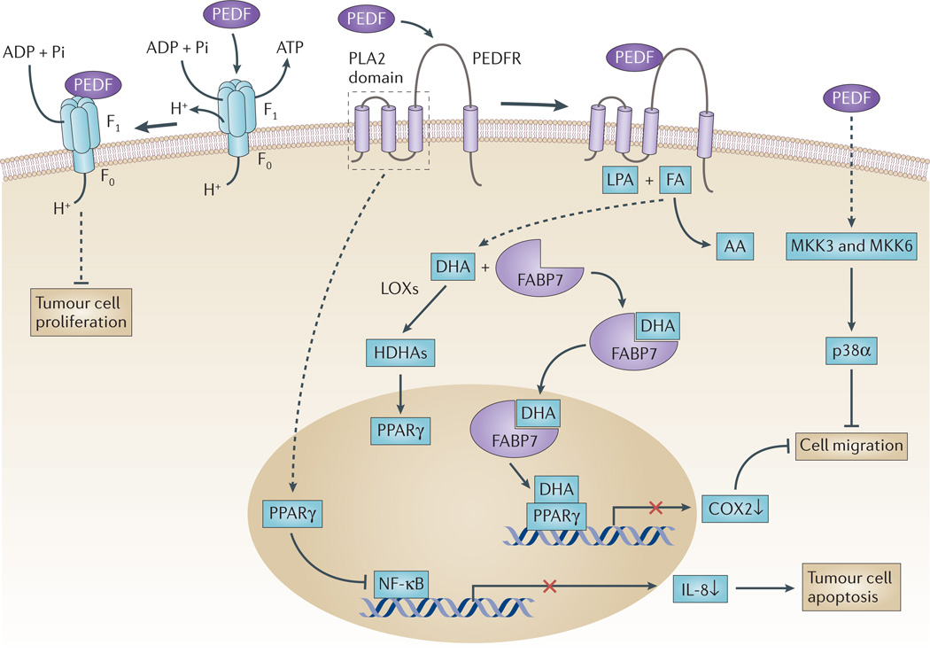
Figure 1. PEDF in tumour cells.
Pigment epithelium-derived factor (PEDF) is a ligand for several receptors, and its interaction with these receptors is thought to trigger the signalling pathways illustrated here. PEDF binds to PED F receptor (PEDFR) and stimulates its phospholipase activity91,151. When PEDFR is at the membrane, its phospholipase A2 (PLA2) active site is located close to the phospholipid bilayer where it can use phospholipids as substrates. Depending on the relative abundance of the fatty acids omega-3 docosahexaenoic acid (DHA) and omega-6 arachidonic acid (AA) in phospholipid membranes, free DHA or AA can be liberated by PEDFR. DHA is a precursor of the anti-angiogenic and neuroprotector neuroprotectin D1 (NPD1)152. Other DHA metabolites, such as hydroxy-DHAs (HDHAs), which are produced by lipoxygenases (LOXs), can act on peroxisome proliferator-activated receptor-γ (PPARγ)102,105,153. Upregulation of PPARγ leads subsequently to the suppression of nuclear factor-κB (NF-κB)-mediated transcriptional activation, reduced production of interleukin 8 (IL-8) and limited proliferation of prostate cancer cells104. Cytosolic fatty-acid-binding protein 7 (FABP7) can bind DHA with higher affinity than AA, translocate DHA to the nucleus and transfer it to PPARγ, thus resulting in the downregulation of promigratory genes, such as cyclooxygenase 2 (COX2)96. PEDF is a ligand of cell-surface F ATP synthase, and its 34-mer peptide region inhibits ATP production and reduces endothelial and tumour cell viability and angiogenesis107,108. PEDF, through interaction with a yet unknown receptor, can sequentially activate MKK3, MKK6 and p38α MAPK to inhibit cell migration77. FA, fatty acid; LPA, lysophosphatidic acid.

