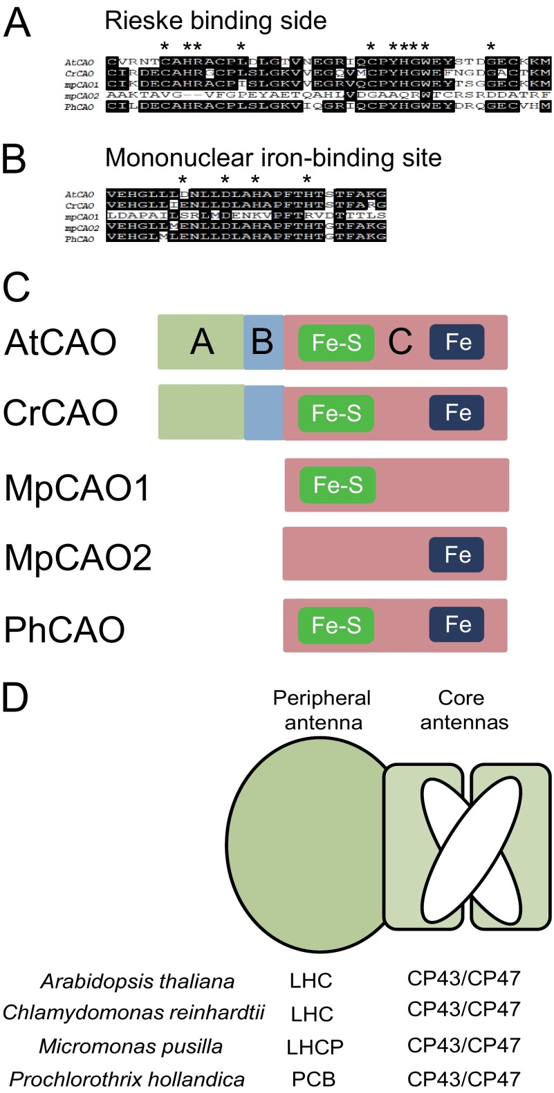
FIGURE 1.
Comparison of the domain structure of the CAO protein among photosynthetic organisms. A, comparison of amino acid sequences of the Rieske center in CAO proteins. B, comparison of amino acid sequences of the mononuclear iron-binding motif in CAO proteins. In both panels, identical residues are shown in white type on a black background. C, domain structure of CAO proteins. Rieske center (Fe-S) and mononuclear iron (Fe)-binding motif are in the C domain. AtCAO, A. thaliana CAO; CrCAO, C. reinhardtii CAO; MpCAO1, M. pusilla CAO (Fe-S); MpCAO2, M. pusilla CAO (Fe); PhCAO, P. hollandica CAO. D, schematic drawing of core/peripheral antenna complexes of Arabidopsis, Chlamydomonas, Micromonas, and Prochlorothrix.

