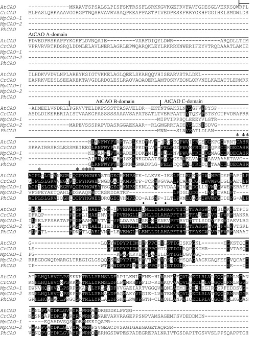
FIGURE 2.
Multiple amino acid sequence alignment of CAO proteins. Identical residues are shown in white type on a black background. The AtCAO sequences corresponding to the A domain, B domain, and C domain are shown. Asterisks and closed squares show binding sites of Rieske center and mononuclear iron-binding motif, respectively.

