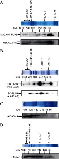
FIGURE 6.
Analysis of the oligomeric states of the CAO proteins. A, two-dimensional BN/SDS-PAGE followed by immunoblotting indicated that MpCAO1 and MpCAO2 proteins accumulated in monomeric, heterodimeric (86 kDa), and higher molecular weight forms in MpCAO1+MpCAO2 plant. B, two-dimensional BN/SDS-PAGE followed by immunoblotting indicated that BCFLAG proteins accumulated in monomeric (43 kDa), trimeric (126 kDa), and higher molecular weight forms in the BCFLAG overexpression plants. The BCFLAG proteins were detected using an anti-FLAG antibody or anti-CAO antibody. C, two-dimensional BN/SDS-PAGE followed by immunoblotting indicated that recombinant AtCAO protein formed higher molecular weight protein complexes in E. coli. The AtCAO proteins were detected using an anti-His antibody. D, two-dimensional BN/SDS-PAGE followed by immunoblotting indicated that PhCAO formed monomeric (41 kDa), trimeric (123 kDa, and higher molecular weight complexes. The PhCAO proteins were detected using an anti-CAO antibody.

