FIGURE 8.
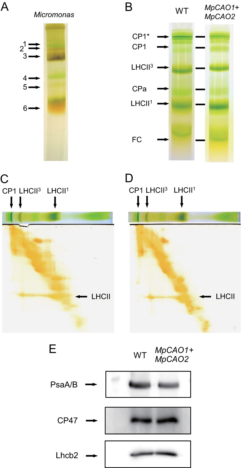
Separation of chlorophyll-protein complexes. Thylakoid membranes of M. pusilla (A), A. thaliana (WT) (B, C, and E), and MpCAO1+MpCAO2 plants (B, D, and E) were prepared. Chlorophyll-protein complexes of those plants were separated by native green gel electrophoresis (A and B). The identity of the chlorophyll-protein complexes corresponding to the bands are presented on the left side of the gel. CP1*, CP1 (PsaA/B)-LHCI complexes; CPa, core complexes of PSII (CP43/CP47); LHCII1 and LHCII3, monomeric and trimeric LHCII, respectively; FC, free Chl. In addition, chlorophyll-protein complexes of WT and MpCAO1+MpCAO2 plants were also analyzed by two-dimensional SDS/SDS-PAGE, followed by silver staining (C and D). The amounts of PsaA/B, CP47, and Lhcb2 proteins were estimated by SDS-PAGE, followed by immunoblot analysis with their specific antibodies (E).

