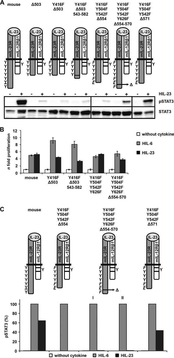
FIGURE 9.
Characterization of the non-canonical STAT3 activation motif in the intracellular domain of IL-23R. A, two IL-23R variants either with a deletion of 17 amino acids (Y416F/Y504F/Y542F/Y626F-Δ554–570) or with the addition of the respective motif (Y416F-Δ503+543-582) were generated and stably transduced into Ba/F3-gp130-mIL-12Rβ1 cells. Resulting Ba/F3 cell lines were washed three times with PBS, starved, and stimulated for 10 min with IL-23. Equal amounts of protein were loaded onto SDS gels. Western blotting was performed using antibodies specific for phospho-STAT3 and STAT3. Data are representative for two experiments. The presented Western blots originate from different membranes and are therefore separated by black lines. B, equal numbers of stably transduced Ba/F3 cells were cultured for 2 days in the presence of 0.2% HIL-6 or 0.2% HIL-23 or without cytokine. Proliferation was measured with the colorimetric CellTiter-Blue® cell viability assay. Values represent the mean value of three repetitions and were normalized. For comparison, n-fold proliferation was calculated by setting the negative control of each Ba/F3 cell line to a value of 1. Data are representative of at least two independent experiments. C, IL-23R variants were washed three times with PBS and starved for 2 h in serum-free DMEM. 1 × 10⁶ cells were stimulated for 30 min with 0.2% HIL-6 or HIL-23, harvested by centrifugation, fixed in 2% (w/v) paraformaldehyde, and permeabilized in 90% (v/v) methanol. Cells were stained for phospho-STAT3 and STAT3 overnight and analyzed by flow cytometry. Two Ba/F3-gp130-mIL-12Rβ1-mIL-23R/Y416F/Y504F/Y542F/Y626F-Δ554–570 cell passages were analyzed, cultivated in the presence of either HIL-6 (I) or HIL-23 (II). Error bars, S.D.

