FIGURE 4.
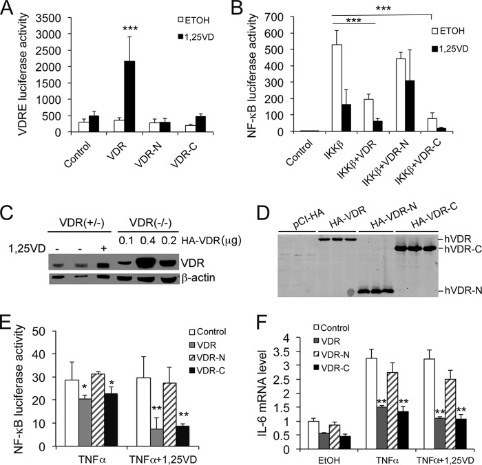
Functional analysis of VDR protein domains in NF-κB regulation. A, VDRE luciferase reporter assays. HEK293 cells were cotransfected with p3xVDRE-Luc and VDR, VDR-N, or VDR-C followed by 24 h of 1,25(OH)2D3 stimulation. ***, p < 0.001 versus the rest. B, effects of hVDR N- and C-terminal fragments on IKKβ-induced NF-κB activity. HEK293 cells were cotransfected with pNF-κB-Luc; IKKβ; and VDR, VDR-N, or VDR-C. The transfected cells were treated with ethanol or 1,25(OH)2D3 followed by luciferase activity assays. ***, p < 0.001. C, comparison of endogenous VDR levels in VDR+/− MEFs and in VDR−/− MEFs transfected with hVDR. VDR+/− MEFs were treated with or without 1,25(OH)2D3 as indicated, and VDR−/− MEFs were transfected with different amounts of HA-hVDR (0.1, 0.2, or 0.4 μg/well) as indicated. Cell lysates were analyzed by Western blot analysis after 24 h using anti-VDR antibodies. Note the comparable VDR levels in VDR+/− MEFs and VDR−/− MEFs transfected with 0.1 μg HA-VDR plasmid/well. D, Western blot analysis with anti-HA antibodies showing that VDR−/− MEFs were reconstituted with empty vector, HA-VDR, HA-VDR-N, or HA-VDR-C by transfection at 0.1 μg plasmid DNA/well. E, VDR−/− MEFs were cotransfected with pNF-κB-Luc and control empty vector, VDR, VDR-N, or VDR-C plasmid (0.1 μg/well). The transfected cells were treated with TNFα or TNFα+1,25(OH)2D3 for 24 h followed by luciferase activity assays. *, p < 0.05; **, p < 0.01 versus corresponding control. F, VDR−/− MEFs were transfected with control empty vector, VDR, VDR-N, or VDR-C (0.1 μg/well). The transfected cells were treated with TNFα or TNFα+1,25(OH)2D3 for 6 h, and the IL-6 transcript was quantified by quantitative PCR. **, p < 0.01 versus corresponding control.

