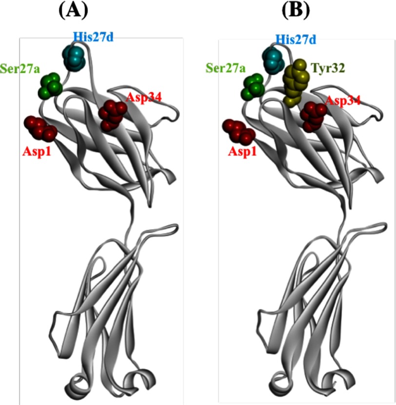
FIGURE 2.
Structure model for 22F6 light chain. A, side chains having the potential to form serine protease-like triads (Asp1 (or Asp34), Ser27a, and His27d) are shown. Asp, His, and Ser are colored red, blue, and green, respectively. The distance between Cα of His27d and Ser27a is 8.26 Å, which is close to the Cα distance (8.42 Å) between them in a catalytic triad of trypsin. B, Tyr32 is visualized. The residue is situated between Asp34 and His27d and may be concerned with the nuclease activity. Models for the light chain were visualized using WebLab ViewerLite (Accelrys Inc., San Diego, CA).

