FIGURE 6.
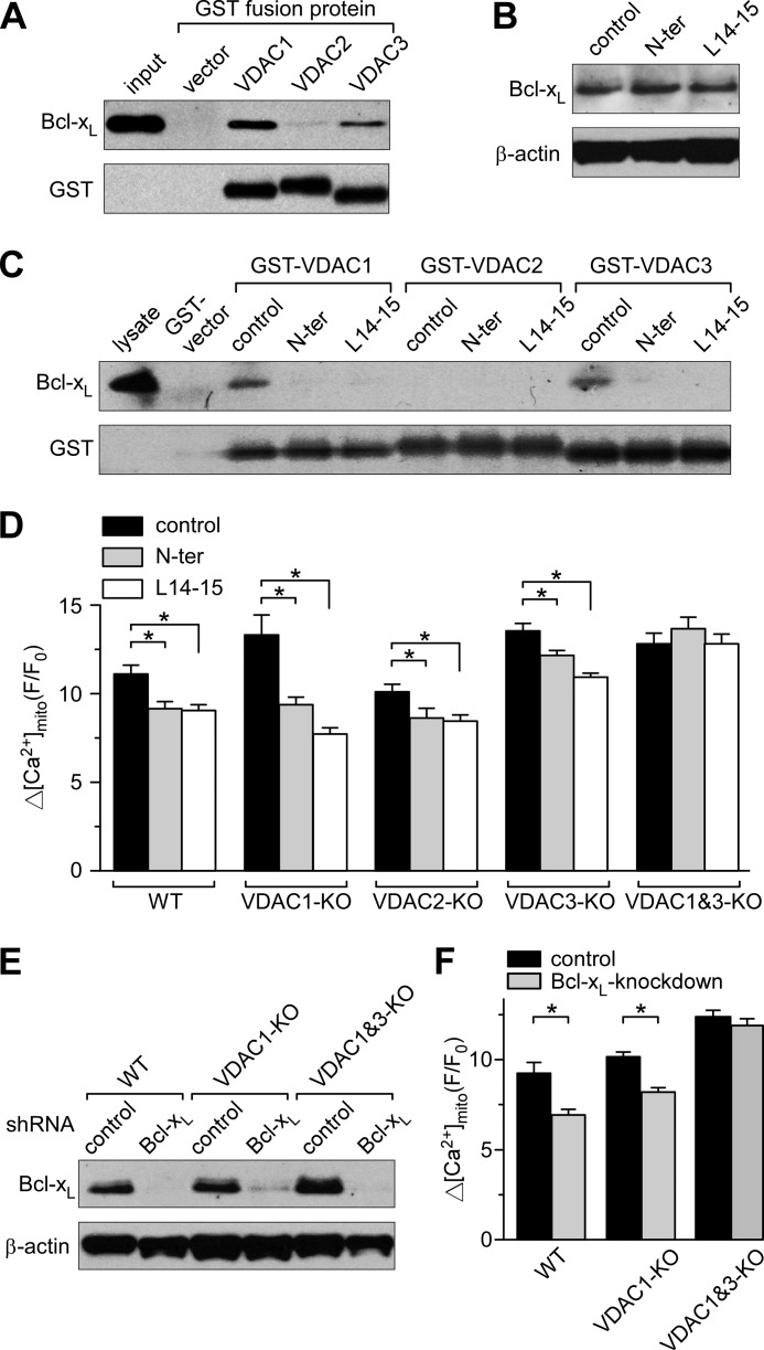
Bcl-xL interacts with VDAC1 and VDAC3 to promote [Ca2+]mito uptake. A, Western blot of recombinant purified Bcl-xL pulled down by GST fusion proteins of VDAC1, -2, and -3 is shown in the upper lanes with the loading control blots of GST depicted below. B, Western blot detecting Bcl-xL expression levels in WT MEF cells after incubation with cell-permeant control peptide or peptides based on the VDAC1 sequence (N-ter and L14-15; 20 μm for 1 h). C, Western blot detection of Bcl-xL pulled down by GST fusion proteins of VDAC isoforms from WT MEF cell lysates pretreated with control or VDAC1-based peptides. D, summary bar graphs showing the peak [Ca2+]mito uptake in permeabilized VDAC knock-out cells stepped from 0 to 3 μm [Ca2+] in the absence or presence of 2 μm VDAC1 peptides (mean ± S.E.; *, p < 0.05; ANOVA). Cell lines included VDAC single knockouts (VDAC1-KO, VDAC2-KO, and VDAC3-KO) and VDAC1 and -3 double knock-out (VDAC1&3-KO). E, Western blot showing Bcl-xL shRNA knockdown in WT, VDAC1 knock-out, and VDAC1 and -3 double knock-out MEF cells. F, summary bar graphs showing the effect of Bcl-xL knockdown on the peak [Ca2+]mito uptake in permeabilized WT and VDAC knock-out cells upon stepping from 0 to 3 μm [Ca2+] (mean ± S.E.; *, p < 0.05; Student's t test). Error bars represent S.E.

