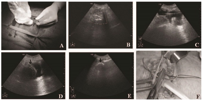
Figure 1.
— (A) Using the ultrasound probe to direct the introducer needle. (B) Doppler ultrasonography shows a localized disturbance just under the peritoneum, because of the flow of peritoneal dialysis fluid into the peritoneal cavity. Deeper vessels can also be delineated. (C) The guidewire is seen in the pelvis, (D) followed by visualization of the catheter coil (black arrows) in the pelvis. (E) Catheter coil seen in another patient. (F) Anterior rectus sheath being closed over the deep cuff by applying a purse-string suture.

