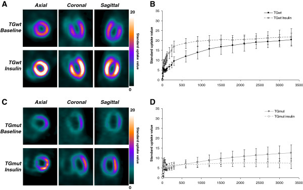
Figure 2.

Representative microPET FDG images and SUV time-activity curves at baseline and following acute insulin treatment. Representative cardiac FDG images of TGwt (A) and TGmut mice (C) at baseline and following insulin stimulation. Images are presented in the axial, coronal, and sagittal views and standardized to the same scale. Myocardial TAC baseline and following insulin in TGwt (B) and TGmut mice (D), respectively, over 60 min using SUV. TGwt n = 3 and TGmut n = 7.
