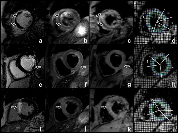
Figure 1.

Patient with MO and IMH (a-d). LGE image demonstrates anterior and septal enhancement, corresponding to scar. The central hypoenhanced core corresponds to MO (a, arrowed). T2w (b) and T2* (c) imaging show central hypoenhancement (arrowed), indicative of IMH. Strain measurements in epicardial, mid-myocardial and endocardial tracks are measured (d) for infarcted (1) and remote (2) zones. A similar arrangement of images for infarction with MO but no IMH is shown (e-h). Note absence of central hypoenhancement in T2w and T2* sequences (arrowed). A patient without MO is shown (i-l).
