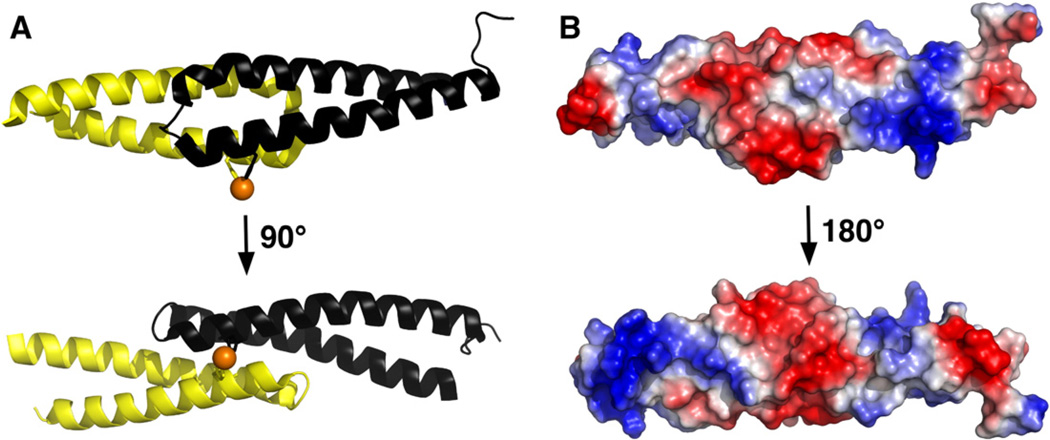
Fig. 1.
(A) Two orientations of the likely cce_0567 dimer in solution that differ by ~90° about the horizontal axis. Each subunit is colored separately and the side chains of the H36 residue from each protein and the nickel (orange) highlighted. (B) The solvent accessible electrostatic surface for the cce_0567 dimer shown in two orientations. The upper orientation is similar to the orientation shown on the top of (A) with the lower orientation differing by ~180° about the horizontal axis.

